
Peripheral Rim Fractures and Occlusal Effect Caries in Teeth
Commonly misdiagnosed as poor hygiene, interproximal decay is often caused by small fractures that expose dentin to bacteria, creating carious lesions. Understanding how occlusal effect caries begins is the first step in prevention and treatment. Early treatment can offer more minimally invasive procedures for patients, conserving tooth structure and long-term health.
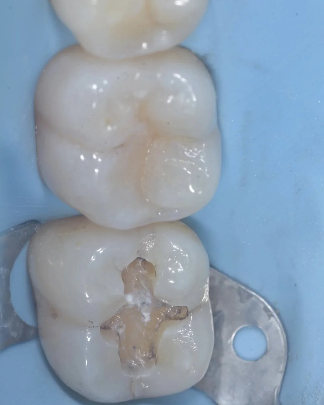
Peripheral rim fractures are small fractures most often found along adjacent contacts and around large restorations. These fractures start in enamel but continue into dentin, increasing the tooth’s risk of developing larger caries and cracks.
Occlusal effect caries is caused by peripheral rim fractures that create a pathway for bacteria to enter into the tooth and create a carious lesion. Occlusal effect caries is distinguished by its symmetric shape around the peripheral rim fracture.


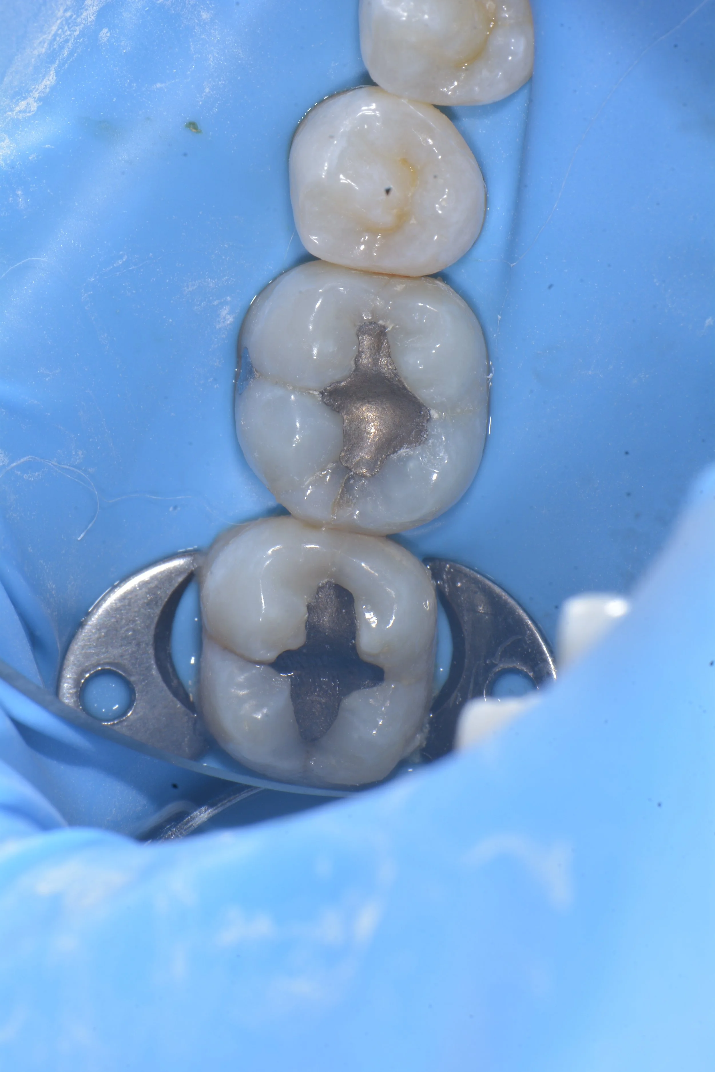
This case by Dr. Davey Alleman, DMD shows how peripheral rim fractures can be treated and the teeth restored with biomimetic protocols to conserve the remaining tooth structure.
What causes peripheral rim fractures?
Peripheral rim fractures develop like any crack in a tooth. As the tooth is stressed over time, molecules can pull apart, initiating a crack. Teeth are naturally designed to withstand cracks thanks to anatomical features like sub-occlusal transverse ridges and flexibility at the dentino-enamel junction (DEJ).
Large restorations are the most common cause of peripheral rim fractures because they remove occlusal anatomy, including sub-occlusal transverse ridges. Without this occlusal buttressing, teeth bend and flex more than they are designed to do during the forces of chewing.
Overloading the tooth in this way increases the likelihood of fracture, which often starts as peripheral rim fractures around the outside of the restoration.
How to diagnose peripheral rim fractures and occlusal effect caries
Peripheral rim fractures are often visible from staining in the crack. Use high magnification of at least 6.5-8x magnification for diagnosis. Carious lesions that are symmetrical around the crack are considered occlusal effect caries and having originated from the peripheral rim fracture. These lesions will stain with caries detector dye but in the initial stages are not visible on x-rays.

These photos by Dr. Davey Alleman, DMD show how a carious lesion forms around a peripheral rim fracture. This lesion is called occlusal effect caries.
Treatment for occlusal effect caries
Treat occlusal effect caries like any carious lesion. Use high magnification and caries detector dye along with the peripheral seal zone concept to achieve caries removal endpoints. Continue with immediate dentin sealing and resin coating to restore the tooth in a way that mimics the form and function of a natural tooth.


How to prevent peripheral rim fractures and occlusal effect caries
The best way to prevent peripheral rim fractures and the occlusal effect caries they cause is to restore teeth in a way that mimics the natural structure of sub-occlusal transverse ridges that prevent cracks from forming in virgin teeth. Bonding at a strength that mimics the tooth’s natural connection to itself (30-50 MPa) allows the restored tooth to bend and flex during the forces of occlusion while resisting crack initiation, even in large restorations. Dr. David Alleman, DDS created his Six Lessons Approach to systematize bonding protocols to dentin, giving practitioners a set of protocols they can use to restore teeth in a way that conserves tooth structure and pulp vitality while mimicking a tooth’s natural form and function. Get trained in the Six Lessons Approach in upcoming Alleman Center dentistry continuing education programs.
Learn more about why dental restorations fail in this Six Lessons Approach Podcast Episode with Dr. David Alleman, DDS.